The Hidden Brain Cells Powering Sleep, Memory, and Disease Defense

Neuroscientist Terrence Sejnowski explains why these long-overlooked support cells may be just as important as neurons — and what they reveal about brain health and disease.
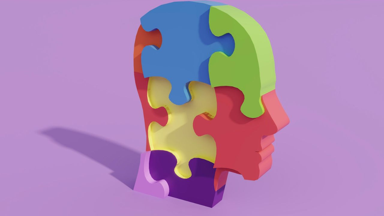
For decades, brain science has centered almost entirely on neurons. But a growing body of research suggests another group of cells has been working behind the scenes to shape everything from how we sleep to how we age — and may even help determine who develops diseases like Alzheimer’s. These “support cells,” known as glia, are emerging as powerful players in brain health.
Terrence Sejnowski, PhD, professor and laboratory head of the Computational Neurobiology Laboratory and Francis Crick Chair at the Salk Institute for Biological Studies, studies how glial cells differ across brain regions and species, and what those patterns might mean for brain function and disease risk.
In a recent Salk study, he and his team compared glial cell organization across different brain regions and mammal species, finding striking patterns in how these cells are arranged. His work looks at how glial cells differ across brain regions and species, and what those patterns might mean for brain function and disease risk.
In a recent Live Talk with Being Patient, Sejnowski explains the three main types of glial cells and their roles. He discusses how glial cells help clean the brain during sleep, how they may be involved early in conditions like Alzheimer’s and multiple sclerosis, and why some research suggests glia could be a better marker of brain health than neurons alone. His insights point to a future in which understanding and protecting glial cells could open new paths for prevention and treatment.
Being Patient: So can you tell us what exactly is a glial cell?
Terrence Sejnowski: The word glia derives from a Greek word meaning glue, and it connects different neurons and vessels. There are three different types. Astrocytes connect the blood vessel to the neurons with nutrients, also important for shaping learning and memory. These are the connections between neurons.
Microglia are really interesting because they’re mobile. They actually go in between the neurons and they gobble up all kinds of detritus that comes out of the neurons and help clean up the mess.
Finally, the third type is the oligodendrocytes. And what they do is they wrap around the axons that the neurons use to communicate and speed up the signaling. So they have very different functions, but they’re all very important.
Being Patient: Microglia are known as the janitors of our brain. And as I understand it, they really only come out in deep sleep. That’s why sleep is so important. Is that true?
Sejnowski: No, they’re always there. They have an important function during deep slow wave sleep, which is to clear out the space between the neurons. For example, especially in Alzheimer’s, the amyloid beta plaque has to get disposed of. But they’re monitors. They go around, and if there’s any damaged part of a neuron or a synapse, a connection between the neurons, they clean it up. You’re right, maybe it’s a janitor job, but without the microglia, you would really be in bad shape; sleep would really be disrupted.
Being Patient: I thought though that that’s why it’s so important to get sleep, because in deep sleep, the microglia are actually cleansing our brains, like purging toxins or what’s not supposed to be there, and that really only happens at nighttime.
Sejnowski: What happens is that the space between the neurons expands, and that then allows a flow of material, and the glia, basically microglia, are there to make sure that it’s going in the right direction, cleaning things out.
Being Patient: Do we know why that happens only in sleep? Do we understand why?
Sejnowski: When you go to sleep, your brain doesn’t stop. It just goes into a completely different mode, and it’s processing information that occurs during the day to help restore and consolidate long-term memories. And that’s part of the process of cleaning things up that maybe you don’t need, things that are maybe even detrimental, like amyloid beta. That’s part of what goes wrong with Alzheimer’s. They get filled with it, and then they die.
“When you go to sleep, your brain doesn’t stop.
It just goes into a completely different mode,
and it’s processing information that occurs
during the day to help restore and
consolidate long-term memories.”
Being Patient: Could you say that glial cells are important to the health of neurons and how they communicate, or what role do they actually play in terms of supporting the structure of the brain?
Sejnowski: The more we studied them, the more we discovered. Initially, they were thought to be nutritive, supportive, and they are. They carry essential nutrients into the neurons from the blood vessels, but they do a lot more.
For example, when the synapses are talking to each other, there’s an astrocyte that has all these little arms and it goes in between all the different synapses. And it helps those synapses communicate by exchanging chemicals, and they’re connected to each other with electrical connections. So they form kind of a basic structure within which the brain operates. It’s really essential.
Being Patient: How do we think about these cells in relation to neurodegenerative diseases? Are they being studied to understand really what goes wrong when somebody first shows signs, maybe when the plaques are forming or while they’re forming? Can they offer us any insight into what’s going wrong in an Alzheimer’s brain, or for that matter, other dementias?
Sejnowski: I have a colleague, Nicola Allen, who actually studies this. Neuroscientists are discovering that they could be involved in the very beginning of Alzheimer’s.
Alzheimer’s, the plaques and tangles, which are seen in the end state when the neurons are dying, occurs way after the early insult that caused it. And there could be many different things that lead to Alzheimer’s. It takes decades to develop, and viruses have been implicated, but there’s a line of research that implicates microglia, and they go rogue. Suppose that you have microglia that are starting to eat good synapses and destroy the connections between neurons. I mean, this is the worst case scenario, but that does happen.
Being Patient: Tell me a little bit about your research, because I know that wasn’t directly, but you looked at several regions of the brain and looked at how, you know, the different functions for glial cells.
Sejnowski: We have a paper that was recently published in the Proceedings of the National Academy of Sciences, and it was the work of two postdoctoral fellows in my lab, Shyam Srinivasan, who’s from India, and Antonio Pino Duarte from Portugal.
What they did is to look carefully and count the number of different types of glia in different brain areas. And they discovered three regularities.
One of the things that was really amazing is that if you look at the density of glial cells — let’s say the number of glial cells in a volume compared to neurons — it varies across different populations. There may be more glia in, for example, one part like the neocortex compared to the cerebellum, which is a motor structure, or the olfactory cortex.
But however, even though it’s different, it’s the same ratio. The density is the same across all mammalian species. And that’s really quite remarkable. What it says is that there’s something that’s characteristic about the glial cells that makes the need for them different in different areas.
Being Patient: How could we use this as a gauge for brain health, perhaps? So your findings are, I guess, suggesting glia may sometimes be a better marker for the brain’s region in certain areas than neurons. What are those next questions that you’re asking in terms of understanding these cells’ relationship to brain function overall?
Sejnowski: Well, a couple of things. First, multiple sclerosis is a devastating disease, a motor disease, and we now know it’s an autoimmune disease, and it’s the oligodendrocytes, the ones that are wrapping the axon, that get attacked by the immune system. And once you start peeling away the oligos, your ability to walk and your ability to move are seriously disrupted. And so clearly, knowing something about what’s going on there and trying to help patients is really important.
Another one is that the astrocytes I mentioned are important for learning and memory. And let me just give you one experiment that I was particularly struck by. Maiken Nedergaard at the University of Rochester did the following experiment. She transplanted astrocytes from humans into rats and mice, rodents. And here’s what she discovered, that the human astrocytes, which are much bigger than the mouse astrocytes, helped the rats learn faster. Okay, well, that’s maybe giving us a hint that these astrocytes are really important for human learning.
There’s a story, which is amazing. When Einstein died, his physician kept his brain, and much later it was studied by neuroanatomists because they figured that, look, this is one of the most amazing brains in physics in terms of him being a genius. Let’s see which parts of the brain were different. They didn’t find any part of the brain that was different in terms of the volume, but what they discovered was that Einstein had more glial cells than the normal person, especially in the part of the brain called the parietal cortex, which is important for your spatial relationships, where you are in space. And he had a wonderful way of visualizing problems and coming up with solutions, like in relativity.
I really think that understanding those mechanisms may help others who are having learning problems, and might be a possible way for us to be able to help people who have learning disabilities.
Being Patient: Is it the relationship between glial cells and neurons that we need to delve into more, or where’s the missing part of the equation in terms of where we are in terms of what we need to know?
Sejnowski: You’re putting your finger on something really important, and that is the communication between different cells in the brain. Now, I mentioned synapses. That’s the very complex way that neurons talk to each other, by releasing a chemical transmitter and then activating the other side, the postsynaptic side.
Well, it turns out that glial cells also release these chemicals. In fact, one of the important functions they have is to take up some of the transmitters, glutamate in particular, and then reprocess it and send it back to the presynaptic side so that it can be recycled. So it’s a recycling system, and it’s incredibly important.
What we’re discovering is that a lot of the signals that are there have multiple functions that have to do with regulating learning, how those changes occur in the synapse that make them more strong in terms of their function, or sometimes weaker.
We know there is something called peptides. These are short chains, like 10 amino acids or less in some cases, that are also released. And the problem is we don’t know what those peptides are doing. And so there’s a big mystery here. And it’s known that the peptides act very slowly in many cases, over many minutes. So they may be having a function in reorganizing the structure of the networks on a longer timescale that may be, again, important for learning and memory, but maybe other functions too, we just don’t know.
Being Patient: We know beta amyloid plaque is the presumed beginning pathology for Alzheimer’s in particular. But is there a hypothesis out there that if we study if there is a problem with the glial cells prior to amyloid, then is that maybe one way where we should look at neurodegeneration? Like maybe they can unlock some of the clues in terms of what goes wrong in an Alzheimer’s brain. Is that one way to put it?
Sejnowski: That’s one hypothesis, and that’s a hypothesis that may be involved in the early triggering. But there is another hypothesis, which is that the microglia are there doing their best. They’re just working full time.
The amyloid beta, there’s been a hundred clinical trials, phase three clinical trials, each costing hundreds of millions and in some cases a billion dollars. They’ve all failed. In other words, just clearing out the amyloid beta is not the solution. The real problem is to go back and figure out what started it. And I gave two examples. The microglia might be involved; they may go berserk. There’s some evidence for that. But also there’s more and more evidence that viruses get in, and that triggers a bunch of changes that then lead to this very end state, which is the plaques.
There was a big study that was done of people who have lived a hundred years. And some of them had amyloid plaques, and they were functioning normally. In other words, it’s not 100 percent that amyloid plaques mean you have Alzheimer’s. So that throws a little bit of doubt on the simple idea that this one particular problem of polymerization of a molecule that makes all these tangles, that’s not the only problem.
NIH now is actually looking for other ways to think about how to attack the problem. It’s getting worse because the population is aging. And it’s a burden, not just on the cost of medical care, but it’s a burden on the families. They’re suffering too because [you have to care for them], 24/7.
“The amyloid beta, there’s been a hundred clinical trials, phase three clinical trials, each costing hundreds of millions and in some cases a billion dollars. They’ve all failed. In other words, just clearing out the amyloid beta is not the solution. The real problem is to go back and figure out what started it.”
Being Patient: Now that you have studied glial cells and realized that across different mammal species we have the same amounts in different parts of the brain, what’s the next step to your research?
Sejnowski: The next step is going to be to try to manipulate. What you can do now with modern genetic techniques, you can manipulate genes, and we can identify genes that may affect the density of these different glial cells. And then, by looking at the impact on behavior and health, that’ll give some hints about what might be their function.